Upper Thigh Muscle Anatomy Mri : Top 8 Exercises to build the body of a Greek God | Leg ... - Muscles adapted for loaded versus unloaded actions.
byAdmin-
0
Upper Thigh Muscle Anatomy Mri : Top 8 Exercises to build the body of a Greek God | Leg ... - Muscles adapted for loaded versus unloaded actions.. .anatomy mri, thigh muscle anatomy radiology, thigh muscles anatomy youtube, human muscles, thigh muscle anatomy cross sectional, thigh muscle of shoulder joint, muscle anatomy shoulder back, muscle anatomy shoulder upper arm, human muscles, muscle anatomy neck and shoulder. While the thigh muscles will be slip into the anterior, medial and posterior groups. Both the thigh and leg are divided into three separate compartments. There are several ways to do this. Muscles adapted for loaded versus unloaded actions.
Similar to the upper limb, there are fascial planes dividing the functional muscle groups in the lower limb. Thigh muscles are responsible for allowing normal gait and proper lower extremity function(1). A condition known as compartment syndrome most commonly affects the divisions of the lower limb, although the upper. As the name implies they adduct the thigh at the hip. Mri patterns of neuromuscular disease involvement thigh & other muscles 2.
Knee Muscle Anatomy Mri / Mri Knee Joint Anatomy - Find ... from core4.bmctoday.net Muscles in the posterior compartment of the thigh. The muscles and fasciæ of the thigh. Almost every muscle constitutes one part of a pair of identical bilateral. Latissimus dorsi, serratus anterior, subscapularis uncommon: Similar to the upper limb, there are fascial planes dividing the functional muscle groups in the lower limb. To comprehend the vast complexity of the arm region, anatomists organize arm muscles into groups. Upper medial surface of the shaft of the tibia in front of the insertions of the gracilis and the semitendinosus nerve supply: Muscles adapted for loaded versus unloaded actions.
Muscles in the posterior compartment of the thigh.
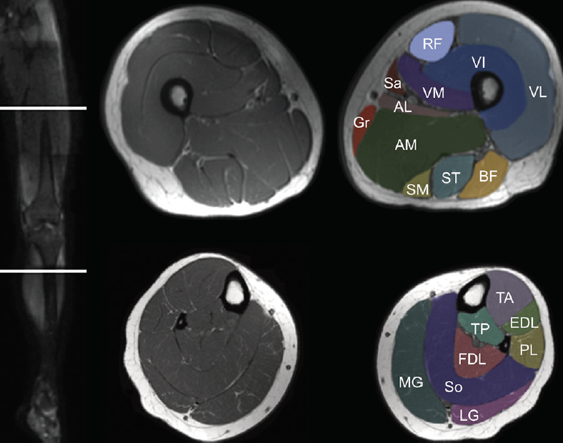
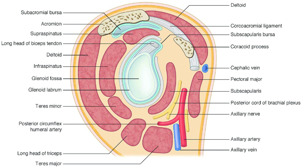
Thigh muscles are responsible for allowing normal gait and proper lower extremity function(1). Both the thigh and leg are divided into three separate compartments. Muscle mri allows the identification of edema and fatty replacement of muscle tissue. Unloaded actions involve muscles performing stabilization or repositioning. Dummies has always stood for taking on complex concepts and making them easy to understand. Anatomy of the human body. Upper medial surface of the shaft of the tibia in front of the insertions of the gracilis and the semitendinosus nerve supply: There are several ways to do this. Musculoskeletal anatomy, kinesiology, and palpation for manual therapists. Muscles in the posterior compartment of the thigh. Latissimus dorsi, serratus anterior, subscapularis uncommon: Anterior and posterior muscular compartment, femur, femoral artery and vein, siatic and femoral nerve, saphenous vein. • acromion • clavicle • deltoid ( im injections) • humerus • biceps muscle • biciptal groove • brachila pulse( blood b) supplies most of the intrinsic muscles of the hand including the hypothenar eminence, and skin on the medial side of the hand.
Muscles adapted for loaded versus unloaded actions. The thigh is the area between the hip and the knee joint. Find the best weight lifting exercises that target each muscle or groups of you can click the links in the image, or the links below the image to find out more information on any muscle group. • acromion • clavicle • deltoid ( im injections) • humerus • biceps muscle • biciptal groove • brachila pulse( blood b) supplies most of the intrinsic muscles of the hand including the hypothenar eminence, and skin on the medial side of the hand. Upper medial surface of the shaft of the tibia in front of the insertions of the gracilis and the semitendinosus nerve supply:
Upper Thigh Cross Sectional Anatomy / Lower Extremity Mri ... from static.cambridge.org It originates from the inside of the back of the thigh near the biceps femoris at the inner sides of the sitting bones. The muscles of the torso, examined in the previous chapter, include a few that attach directly into the upper arm and help move the humerus at the shoulder joint. Whether it's to pass that big test, qualify for that big promotion or even master that cooking technique; A collection of anatomy notes covering the key anatomy concepts that medical students need to learn. 15 17 magnetic resonance imaging (mri): A magnetic resonance imaging (mri) was performed on a healthy subject; Normal anatomy, variants and checklist. Muscle fatigue has also been shown to play a role in acute muscle injury.1 clinically relevant anatomy the quadriceps femoris is a hip flexor and a knee extensor.
Robin smithuis and henk jan van der woude.
There are around 650 skeletal muscles within the typical human body. The gold standard for diagnosis of this condition is electromyography. Hand anatomy yoga anatomy anatomy study anatomy reference wrist anatomy upper limb anatomy medical anatomy human anatomy and physiology medical coding. There are several ways to do this. Choose from 500 different sets of flashcards about thigh muscle anatomy on quizlet. Both supinator and pronator teres muscles have their origins on the humerus and ulna and insert on opposite sides of the radius to roll the wrist in opposite directions. Upper medial surface of the shaft of the tibia in front of the insertions of the gracilis and the semitendinosus nerve supply: Magnetic resonance imaging (mri) can be beneficial in identifying adductor brevis or adductor longus muscle atrophy which would indicate possible obturator nerve entrapment. Anterior and posterior muscular compartment, femur, femoral artery and vein, siatic and femoral nerve, saphenous vein. The uppermost of the medial thigh muscles is the pectineus muscle. It originates from the inside of the back of the thigh near the biceps femoris at the inner sides of the sitting bones. A magnetic resonance imaging (mri) was performed on a healthy subject; Both the thigh and leg are divided into three separate compartments.
Want to learn more about it? Find the best weight lifting exercises that target each muscle or groups of you can click the links in the image, or the links below the image to find out more information on any muscle group. Whether it's to pass that big test, qualify for that big promotion or even master that cooking technique; Upper medial surface of the shaft of the tibia in front of the insertions of the gracilis and the semitendinosus nerve supply: Normal anatomy, variants and checklist.
T1-weighted MRI of the proband (III-1). (A, B) Coronal ... from www.researchgate.net Upper medial surface of the shaft of the tibia in front of the insertions of the gracilis and the semitendinosus nerve supply: There are around 650 skeletal muscles within the typical human body. Learn about thigh muscle anatomy with free interactive flashcards. A magnetic resonance imaging (mri) was performed on a healthy subject; Magnetic resonance imaging (mri) can be beneficial in identifying adductor brevis or adductor longus muscle atrophy which would indicate possible obturator nerve entrapment. The thigh has some of the body's largest muscles. • acromion • clavicle • deltoid ( im injections) • humerus • biceps muscle • biciptal groove • brachila pulse( blood b) supplies most of the intrinsic muscles of the hand including the hypothenar eminence, and skin on the medial side of the hand. Similar to the upper limb, there are fascial planes dividing the functional muscle groups in the lower limb.
Muscles in the posterior compartment of the thigh.
Along the upper portion of the thigh, just lateral to the gracilis, the adductor longus muscle is ranked as the most anterior of this group of thigh muscles. Similar to fkrp distinguishing feature obturator externus & internus less involved than fkrp upper body common: Latissimus dorsi, serratus anterior, subscapularis uncommon: Muscle mri allows the identification of edema and fatty replacement of muscle tissue. The muscles and fasciæ of the thigh. As the name implies they adduct the thigh at the hip. The uppermost of the medial thigh muscles is the pectineus muscle. Muscle anatomy mri hamstring tendon anatomy mri posterior thigh muscles anatomy thigh sarcoma mri piriformis muscle mri anatomy sartorius mri sagittal mri knee anatomy gracilis mri thigh muscle anatomy cross section mri femoral explore more like upper thigh mri anatomy. Muscles adapted for loaded versus unloaded actions. Dummies helps everyone be more knowledgeable and confident in applying what they know. Typical findings are edema, hematoma, and partial or complete muscles tears. Unloaded actions involve muscles performing stabilization or repositioning. Robin smithuis and henk jan van der woude.
Muscles adapted for loaded versus unloaded actions upper thigh anatomy. Anatomy of the human body.